Monday, November 23, 2009
Saturday, July 4, 2009
Rutherford's Nuclear Model
After the Geiger–Marsden experiment (also called the Gold foil experiment or the Rutherford experiment), conducted by Rutherford, and his students Hans Geiger and Ernest Marsden, by bombarding alpha particles on a very thin gold foil, Rutherford concluded that:
- Most of the space in the atom is empty
- The positive charge and most of the mass of the atom was concentrated in a small volume called nucleus
- The size of the nucleus (10-15) is very small compared to that of the atom (10-10)
- Electrons move around the nucleus with very high speed in circular paths called orbits
- Electrons and nucleus are held together by electrostatic forces of attraction
Radioactivity and Radioactive Elements
Henri Becqueral observed that certain elements emit radiation on their own. This phenomenon is known as radioactivity. And such elements are called radioactive elements.
Other scientists in this field include Marie Curie, Piere Curie, Rutherford and Fredrick Soddy.
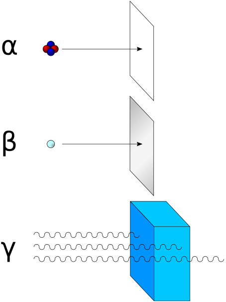
α-rays = 2 protons +2 neutrons
β-rays = -ve particles (electron or positron)
γ-rays are electromagnetic radiatons of very high energy (do not contain particles)
α-rays have the least penetrating power. β-rays have a penetrating power 100 times that of α-rays.
And γ-rays have 1000 times that of α-rays

Other scientists in this field include Marie Curie, Piere Curie, Rutherford and Fredrick Soddy.
α-rays = 2 protons +2 neutrons
β-rays = -ve particles (electron or positron)
γ-rays are electromagnetic radiatons of very high energy (do not contain particles)
α-rays have the least penetrating power. β-rays have a penetrating power 100 times that of α-rays.
And γ-rays have 1000 times that of α-rays
X-rays
When electrons strike a material in the CRT, rays are produced which can cause fluorescence in fluorescent materials. These were called X-rays by its discoverer (Wilhalm Roentgen)(1895)
X-rays are produced effectively when electrons strike dense metal anode (called targets). These are not reflected by electric or magnetic fields (but they possess electro-magnetic character) and have very high penetrating power.
Wavelength = ~0.1 nm
X-rays are produced effectively when electrons strike dense metal anode (called targets). These are not reflected by electric or magnetic fields (but they possess electro-magnetic character) and have very high penetrating power.
Wavelength = ~0.1 nm
Thomson Model of Atom
JJ Thomson proposed in 1898 that atom was spherical (radius approximately 10-10 m). That the positive charge was uniformly distributed in this sphere. And that the electrons were embedded in it like the plum in pudding, in such a manner that the atom is in a stable electrostatic state.
It is called plum pudding, raisin pudding or watermelon model.
Mass of the atom was assumed to be uniformly distributed over the atom (Rutherford said it to be localized in nucleus)
Thomson got Nobel in 1906 not for this no non-sense model (which not only helped in explaining the neutrality of the atom, but is also accepted as a standard for computational models involving spherical systems like golf balls, global weather models, and biological viruses), but for his investigations on the conduction of electricity by gases.
Neutron
Discovered by Chadwick (1932) by bombarding a thin sheet of beryllium (Be) by alpha particles. Thus neutral particles of mass slightly greater than protons (neutrons) were emitted.
Protons
Canal rays (rays containing positively charged particles) :
- depend upon the nature of the gas present in the CRT. These are the positively charged ions (so, will have as many protons as the gas in the tube)
- charge to mass ratio depends on gas
- some particles carry a multiple of the fundamental unit of electrical charge.
- opposite behaviour to cathode rays in electrical or magnetic field (charge different)
Monday, June 29, 2009
Electron
In 1830, Michael Faraday showed that when electricity is passed through a solution of an electrolyte, chemical reactions occurred at the electrodes, which resulted in the liberation and deposition of matter at the electrodes. This suggested the particulate nature of electricity.
A Crookes tube is an early experimental electrical discharge tube, invented by British physicist William Crookes. It consists of a partially (but not completely) evacuated glass cylinder of various shapes, with two metal electrodes at either end. When a high voltage is applied between the electrodes, current starts flowing through a stream of particles traveling in straight lines from the cathode to the anode. These were called cathode rays or cathode ray particles.
If the anode is perforated, and the anode end of the tube is coated with phosphorescent material zinc sulphide, a bright spot on the coating is developed (which is the principle in tv sets too).
In 1897, J J Thomson (British physicist) measured the ratio of electrical charge (e) to the mass of electron (me) by using CRT and applying electrical and magnetic field perpendicular to each other as well as to the path of electrons. Thomson argued that the amount of deviation of the particles from their path in the presence of electrical or magnetic field depends upon:
e/me = 1.758820 × 10¹¹ C kg -1
where me is the mass of the electron in kg and e is the magnitude of the charge on the electron in coulomb (C). Since electrons are negatively charged, the charge is -e.
Charge on electron:
RA Millikan devised oil drop experiment to determine the charge.
He found −1.6×10⁻¹⁹ C as the value. The present value is −1.6022×10⁻¹⁹ C.
The mass of the electron therefore is
me = e/(e/me) = −1.6022×10⁻¹⁹ C / 1.758820 × 10¹¹ C kg -1 = 9.1094 × 10⁻³¹ kg
A Crookes tube is an early experimental electrical discharge tube, invented by British physicist William Crookes. It consists of a partially (but not completely) evacuated glass cylinder of various shapes, with two metal electrodes at either end. When a high voltage is applied between the electrodes, current starts flowing through a stream of particles traveling in straight lines from the cathode to the anode. These were called cathode rays or cathode ray particles.
If the anode is perforated, and the anode end of the tube is coated with phosphorescent material zinc sulphide, a bright spot on the coating is developed (which is the principle in tv sets too).
- Cathode rays start from cathode (-ve) and move towards anode (+ve)
- These are not visible themselves. But fluorescent and phosphorescent materials glow when hit by them.
- These rays travel in straight lines in the absence of electrical or magnetic field.
- In their presence, they travel like negatively charged particles (suggesting that these contain -ve particles, electrons)
- The characteristics of cathode rays do not depend upon the material of electrodes and the nature of the gas present in the cathode ray tube.
In 1897, J J Thomson (British physicist) measured the ratio of electrical charge (e) to the mass of electron (me) by using CRT and applying electrical and magnetic field perpendicular to each other as well as to the path of electrons. Thomson argued that the amount of deviation of the particles from their path in the presence of electrical or magnetic field depends upon:
- the magnitude of -ve charge on the particle (directly proportional to deflection)
- mass of the particle (inversely proportional to deflection)
- the strength of the electrical or magnetic field (directly proportional to the deflection)
e/me = 1.758820 × 10¹¹ C kg -1
where me is the mass of the electron in kg and e is the magnitude of the charge on the electron in coulomb (C). Since electrons are negatively charged, the charge is -e.
Charge on electron:
RA Millikan devised oil drop experiment to determine the charge.
He found −1.6×10⁻¹⁹ C as the value. The present value is −1.6022×10⁻¹⁹ C.
The mass of the electron therefore is
me = e/(e/me) = −1.6022×10⁻¹⁹ C / 1.758820 × 10¹¹ C kg -1 = 9.1094 × 10⁻³¹ kg
Structure of Atom
The existence of atoms has been proposed since the time of early Indian and Greek philosophers (400 BC). It was thought to be indivisible and therefore called atom derived from the Greek word 'a-tomio' which means uncut-able or non-divisible
John Dalton (British school teacher) proposed atomic theory of matter on scientific basis in 1808.
John Dalton (British school teacher) proposed atomic theory of matter on scientific basis in 1808.
Sunday, June 28, 2009
Difference between Plant cells and Animal Cells
Plant Cell | Animal Cell | |
Cell wall is the outermost layer | Cell membrane is the outermost layer | |
Has definite shape | Does not have definite shape | |
Plastids are present | No plastids | |
No centrioles | Centrioles are present | |
vacuoles are large (may even be 95% of the cell volume) and many | vacuoles are small, few or absent | |
Difference between Eukaryotic cells and Prokaryotic cells
Eukaryotes | Prokaryotes |
Large in size (10 µm to 20 µm) | Small in size (1 µm to 10 µm) |
Outer covering –
| Outer covering is the cell envelope |
Membrane bound cell organelles are present | There are no membrane bound cell organelles |
Have true nucleus with nuclear membrane, chromatin reticulum, and nucleolus | No true nucleus. Instead there is the nucleoid |
Cell divides mainly by mitosis and meiosis | Cell divides mainly by amitosis |
Eukaryotic Cells
includes all
Possess an organised nucleus with nuclear envelope.
Have a variety of complex locomotory and cytoskeletal structures.
Genetic material is organised into chromosomes.
There is also the ribosomes which are present in prokaryotes as well.
- protists
- plants
- animals
- fungi
- the endoplasmic reticulum (ER)
- the golgi complex
- lysosomes
- mitochondria
- microbodies
- vacuoles
Possess an organised nucleus with nuclear envelope.
Have a variety of complex locomotory and cytoskeletal structures.
Genetic material is organised into chromosomes.
There is also the ribosomes which are present in prokaryotes as well.
Range and Order of Lengths
The sizes of the objects in the universe varies in a wide range.
Size of a proton | 10-15 |
Size of atomic nucleus | 10-14 |
Size of hydrogen atom | 10-10 |
Length of typical virus | 10-8 |
Wavelength of light | 10-7 |
Size of RBC | 10-5 |
Thickness of a paper | 10-4 |
Height of the Mt. Everest above sea level | 104 |
Radius of the Earth | 107 |
Distance of moon from the earth | 108 |
Distance of the Sun from the earth | 1011 |
Distance of Pluto from the Sun | 1013 |
Size of our galaxy | 1021 |
Distance to Andromeda galaxy | 1022 |
Distance to the boundary of observable universe | 1026 |
There are also some special units for large and small lengths
1 fermi = 1 f = 10-15 m
1 angstrom = 1 Å = 10-10 m
1 astronomical unit = 1 AU (average distance of the Sun from the Earth) = 1.496 × 10¹¹ m
1 light year = 1 ly = 9.46 × 10¹⁵ m (distance that light travels at 3 × 10⁸ m/s in a year)
1 parsec = 3.08 × 10¹⁶ m (distance at which average radius of earth's orbit subtends an angle of 1 arc second)
Physics home
Measurement of Length
For measuring large distances, parallax method is used
Parallax is an apparent displacement of an object viewed along two different lines of sight.
If we hold a pencil such that it covers a spot on the wall when our left eye is closed, and then open the left eye and close the right, parallax can be observed.
The distance between the two points of observation is called basis.
If we observe a planet S at a distance D from earth, from two points A and B which are d distance apart on earth, then, angle inscribed by AB on the planet, angle ASB is called parallax angle or parallactic angle. Here, we take AB as an arc of the circle with centre S and radius D.
So, D=b/angle ASB (if angle ASB is taken in radians). Thus we get D. (basis/angle)
Similarly, we can determine the diameter of the planet. If d is the diameter, and dAd' the angular size (angle subtended by d at earth),
then dAd' = d/D (diameter / distance)
For estimation of very small distances:we will have to use special methods.
An optical microscope can view objects of size greater than its wavelegth (4000 Å to 7000 Å) 1 angstrom = 1.0 × 10⁻¹⁰ meters.
For smaller wavelengths, electron beams are used (electron micrsoscope). But due to the wave behaviour of electrons even this method has limitations. The maximum resolution of electron microscope is upto 0.6 Å.
Tuunneling microscopy is being developed which can go to even smaller scales.
But, the size of molecules can be estimated.
Oleic acid is a soapy liquid with a large molecular size.
- 1 cu.cm of oelic acid is dissolved in alcohol to get 20 cu.cm solution.
- 1 cu.cm of this solution is taken and diluted to 20 cu.cm using alcohol
Now, the concentration of the solution is 1/400 cu.cm of oelic acid per cu.cm - some lycopodium powder is sprinkled on surface of water in a large trough
- one drop of this solution is put in the water
the oelic acid drop spreads into a thin, large, circular film of molecular thickness - The diameter of the film is measured to get its area.
- The volume of the amount of drops we've put is determined.
Let that be n cu.cm - The amount of oelic acid in this solution is
n/400 cu.cm - The oelic acid forms a thin layer of thickness t
this t = volume of the film/area of the film
ie t = n / (400 * area ) cm - if the film is of mono-molecular thickness, then this becomes the diameter of oelic acid molecule.
Physics home
The Characteristics of a Good Unit
A good unit:
- Must be internationally accepted
- Must be well-defined
- Must not vary with physical conditions, place or time
- Must be of a size not too large or too small compared to the physical quantity that is to be measured
- Must be accurate
- Must be easily accessible
- Must be easily reproducible
- The conversions within a system of units must be simple and convenient
Physics home
The International System of Units
Earlier, different countries were using different systems of measurement like CGS, FPS (or British) and MKS system
- CGS: centimetre, gram, second
- FPS: foot, pound, second
- MKS: metre, kilogram, second
Now, there is an internationally accepted system of measurements known as Systeme Internationale d' Unites (French for International System of Units) abbreviated as SI. It was developed by General Conference on Weights and Measures in 1971 for international usage.
Length | metre | m | Distance travelled by light in vacuum during 1/299,792,458 second |
Mass | kilogram | kg | Mass of international prototype of kilogram at international Bureau of Weights and Measures |
Time | second | s | duration of 9,192,631,770 periods of radiation corresponding to the transition between the two hyperfine levels of the ground state of cesium-133 atom |
Electric current | ampere | A | that constant current which if maintained between two straight parallel conductors of infinite length, placed 1 metre apart in vacuum would produce a force 2 × 10⁻⁷ newton per metre of length |
Thermodynamic temperature | Kelvin | K | 1/273.16 of the thermodynamic temperature of the triple point of water |
Amount of substance | mole | mol | amount of substance of a system which contains as many elementary entities as there are atoms in 0.012 kilogram of carbon-12 |
Luminous intensity | candela | cd | luminous intensity in a given direction of a source that emits monochromatic radiation of frequency 540 × 1012 hertz and that has a radiant intensity in that direction of 1/683 watt per steradian |
When mole is used elementary entities must be specified.
Units and Measurement
Physical Quantity: Anything that can be measured directly or indirectly.
Fundamental or Base Quantities: Basic non-derivable quantities (length, mass, time, electric current, thermodynamic temperature, amount of substance and luminous intensity)
Derived quantities: Quantities which are obtained from one or more fundamental quantities eg: velocity, acceleration
Supplementary Quantities: eg: solid angle, plane angle
Unit is the reference standard used to measure physical quantity.
Fundamental units: The reference standard used to measure fundamental physical quantities. (metre (m), kilogram (kg), second (s), ampere (A), Kelvin (K), mole (mol), candela(cd))
Derived units: The reference standard used to measure derived physical quantities. Eg newton, pascal, m/s
Supplementary units: The reference standard used to measure supplementary physical quantities. (for plane angles, radian, for solid angle – steradian) radian=length of arc/radius, steradian = intercepted area of sphere/radius
Nucleoid
Nucleoid is an irregular shaped clear region in the cytoplasm of prokaryotic cells which has nuclear material without a nuclear membrane and where the genetic material is localized.
The genome of prokaryotic organisms generally is a circular, double-stranded piece of DNA, of which multiple copies may exist at any time.
A genophore is the DNA of a prokaryote.This is commonly referred to as a prokaryotic chromosome. The term chromosome is misleading for a genophore because the genophore lacks chromatin
Plasmid and episome
A plasmid is an extra-chromosomal DNA molecule separate from the chromosomal DNA which is capable of replicating independently of the chromosomal DNA.
An episome is a portion of genetic material that can exist independent of the main body of genetic material (called the chromosome) at some times, while at other times is able to integrate into the chromosome.
Examples of episomes include insertion sequences and transposons. Viruses are another example of an episome. Viruses that integrate their genetic material into the host chromosome enable the viral nucleic acid to be produced along with the host genetic material in a nondestructive manner. As an autonomous unit (i.e., existing outside of the chromosome) however, the viral episome destroys the host cell as it commandeers the host's replication apparatuses to make new copies of itself.
Another example is an hfr cell (also called hfr strain) which is a bacterium with a conjugative plasmid (often the F-factor) integrated into its genomic DNA. Hfr is the abbreviation for high frequency recombination
An episome is distinguished from other pieces of DNA that are independent of the chromosome (i.e.,plasmids) by their large size.
Plasmids are different from episomes, as plasmid DNA cannot link up with chromosomal DNA. The plasmid carries all the information necessary for its independent replication. While not necessary for bacterial survival, plasmids can be advantageous to a bacterium. For example, plasmids can carry genes that confer resistance to antibiotics or toxic metals, genes that allow the bacterium to degrade compounds that it otherwise could not use as food, and even genes that allow the bacterium to infect an animal or plant cell. Such traits can be passed on to another bacterium.
Inclusion Bodies
Reserve material in prokaryotic cells are stored in the cytoplasm in the form of inclusion bodies. These are not bound by any membrane system and lie freely in the cytoplasm. Eg:
- Phosphate granules
- Sulphur granules
(The phosphate granules and sulphur granules are together called metachromatic granules because when they are treated with dyes they appear as coloured) - Cyanophycean granules
- Glycogen granules
- Gas vacuoles are found in blue green and purple and green photosynthetic bacteria
Prokaryotic Ribosome
In prokaryotes, ribosomes are associated with the plasma membrane. They are about 15 nm by 20 nm in size (nanometre = 10-9 m).
These are granular structures composed of ribonucleic acid (RNA) and proteins, and not surrounded by any membrane.
Made of two subunits
- 50S unit
- 30S unit
But it adds up to 70S. Here, S is the Svedberg's unit and stands for the sedimentation coefficient which is indirectly a measure of density and size.
The Eukaryotic ribosomes are similar but are 80S and have subunits 60S and 40S. But chloroplasts and mitochondria in eukaryotes have 70S ribosomes.
George Palade first observed ribsomes under electron microscope as dense particles in 1953
Ribosomes are the site of protein synthesis.
Sometimes, several ribosomes may attach to a single mRNA and form a chain called polyribosomes or polysome. These ribosomes translate the mRNA into proteins.
Cell Envelope and its modifications
Most prokaryotic cells, especially bacteria have chemically complex cell envelope which consists of a tightly bound 3 layered structure.
- Glycocalyx (outermost)
- Cell wall
- Plasma membrane
Though each has different functions, all of them act together as a protective unit.
Bacteria can be
- Gram positive (those that take up gram stain) (contains teichoic acid) (eg: Staphylococci, Streptococci, Bacillus, Clostridium) [see wikipedia article about cell wall of Gram +ve]
- Gram negative (those that do not take up gram stain) (contains Lipopolysaccharides (LPS), also known as lipoglycans) (eg: Escherichia coli, Salmonella, Shigella) [See wikipedia article about cell wall of Gram -ve]
on the basis of differences in the cell envelopes and the manner in which they respond to the staining procedure developed by Gram.
Glycocalyx differs in composition and thickness. It actually is a network of polysaccharides that project from cellular surfaces. It can be:
- Slime layer (a loose sheath) (The function of the slime layer is to protect the bacteria cells from environmental dangers such as antibiotics and desiccation). (autoclaving or flushing with boiling water are the only certain methods of decontamination)
- Capsule (thick and tough) (prevents phagocytosis, ie engulfing solid particles by the cell membrane to form an internal phagosome)
Cell wall determines the shape of the cell and provides structural support (prevents bursting or collapsing) It is made up of peptidoglycan (also called murein)
Plasma Membrane is semi-permeable and interacts with outside world. It is similar to eukaryotic plasma membrane
The mesosome is a special membraneous structure formed by the extension of plasma membrane into the cell. They may be in the form of
- Vesicles
- Tubules
- Lamella
They help in
- Cell wall formation
- DNA replication and distribution to daughter cells
- Respiration
- Secretion
- Increasing surface area of plasma membrane and enzymatic content
(But actually whether they perform all these functions is in doubt [:D])
In some prokaryotes like cyanobacteria, there are other membraneous extensions called chromatophores that contain pigments. (Chromatophores are responsible for skin colouring in cold blooded animals too)
Motility:
Bacteria may be motile or non-motile. If motile, they will have thin filamentous extensions from the cell wall called flagella.
Flagellum : is different from eukaryotic flagellum. Is composed of three parts:
- Basal body (embedded in cell envelope)
- Hook (embedded in cell envelope)
- Filament (longest portion and extends from cell surface to the outside)
Based on the arrangement of flagella there can be bacteria of type:
- Monotrichous : single flagellum at one end
- Lophotrichous: many flagella at one end
- Amphitrichous : flagella at both ends
- Peritrichous: flagella all over the body.
Besides flagella there are structures, that do not play a role in motility:
- Pili: elongated tubular structures made of a special protein (oligomeric pilin proteins)
(Pili connect a bacterium to another of its species, or to another bacterium of a different species, and build a bridge between the cytoplasms of the cells. This enables the transfer of plasmids between the bacteria. An exchanged plasmid can code for new functions, e.g., antibiotic resistance.)
(During bacterial conjugation, a sex pilus emerging from one bacterium ensnares the recipient bacterium, draws it in, and eventually triggers the formation of a mating bridge, which establishes direct contact, merging the cytoplasms of two bacteria via a controlled pore. This pore allows for the transfer of bacterial DNA from the bacteria with the pilus (donor) to the recipient bacteria. Through this mechanism of genetic transformation, advantageous genetic traits can be disseminated amongst a population of bacteria. Not all bacteria have the ability to create sex pili, however sex pili can form between bacteria of different species.) - Fimbriae: small bristle like fibres sprouting out of the cell. (helps bacteria attach to rocks in streams or to host tissues) (these are actually small pili that help to attach bacteria together when the pili is used for connecting)
Prokaryotic Cell
Eg:
- bacteria,
- cyanobacteria,
- mycoplasma (PPLO: Pleuro Pneumonia Like Organisms – discovered by Edmond Nocard)(lack a cell wall, so is pleomorphic, ie, has the ability to change shape, Without a cell wall, they are unaffected by many common antibiotics such as penicillin or other beta-lactam antibiotics that target cell wall synthesis.)
- rickettsiae (has no cell wall, so, is a pleomorphic organism too) (The majority of Rickettsia bacteria are susceptible to antibiotics of the tetracycline group.)
Rickettsia species are carried as parasites by many ticks, fleas, and lice, and cause diseases such as typhus, rickettsialpox, Boutonneuse fever, African Tick Bite Fever, Rocky Mountain spotted fever, Australian Tick Typhus, Flinders Island Spotted Fever and Queensland Tick Typhus [2] in human beings.
They have also been associated with a range of plant diseases.
Like viruses, they only grow inside living cells
Prokaryotes are smaller than eukaryotes, and multiply more rapidly than eukaryotes.
Bacteria can be of four basic shapes
- bacillus (rod like)
- coccus (spherical)
- vibrio (comma shaped)
- spirillum (spiral)
But still the organization of all prokaryotic cells is similar.
All prokaryotes have
- cell wall surrounding cell membrane (forming the cell envelope)
- cytoplasm
- no well defined nucleus (naked genetic materials).
Bacteria have a small circular DNA outside the genomic DNA (the single chromosome/circular DNA). These smaller DNA are called plasmids. The plasmid DNA decides phenotypic (observable) characters of the bacteria such as resistance to antibiotics. It is also used for monitoring bacterial transformation with foreign DNA. - Ribosomes are the only organelles of eukaryotes that are present in prokaryotes too.
- Inclusions
- Mesosome (specialised differentiated form of cell membrane – infoldings of cell membrane)
Saturday, June 27, 2009
An overview of cell
Eukaryotes have membrane bound distinct strctures called organelles like endoplasmic reticulum (ER), the golgi complex, lysosomes, mitochondria, microbodies, and vacuoles.
The non-membrane bound organelles found in eukaryotes and prokaryotes are Ribosomes (found in cytoplasm, chloroplasts (in plants), mitochondria, rough ER)
The non-membrane bound organelle found in animal cell is centriole which helps in cell divison
(so, animal cell has 2 non-membrane organelle, plant cell -1)
Cells differ in size, shape, activities
Bacteria could be 3 to 5 µm.
Mycoplasmas the smallest cells are only 0.3 µm long. ("Mycoplasmas the smallest cells are only 0.3 µm (micrometre) long.") (1 µm = 0.000001 m = 1×10⁻⁶ m)
The egg of an ostrich is the largest isolated single cell
RBC = 7.0 µm.
Nerve cells are the looooooooooooooooooooooonnnnnnnnnnnnngest cells
There may be disc like, polygonal, columnar, cuboid, thread like or even irregular cells. The shape vary with function
Contributions of Different Scientists
Anton von Leeuwenhoek first observed and described a live cell. (Robert Hook observed cork cell which was dead)
Robert Brown discovered the nucleus.
And all this and future discoveries were only due to the invention of microscope or anything in between simple microscope and the latest electron microscope
Matthias Schleiden (German botanist) examined plants and saw that all plants were composed of different cells that form tissues. (Sch'l'eiden and P'l'ant) (1838)
Theodore Schwann (British as in text, actually German zoologist) studied animal cells and found that cells had a thin outer layer (which is today called plasma membrane). He observed plant cells and found that cell wall was a unique character of plant cells. (1839) (Schw'ann' and 'an'imals)
Thus was put forward the hypothesis that the bodies of plants and animals are composed of cells and cell products.
Thus, Schleiden and Schwann together formulated the cell theory. But didn't explain how new cells were formed.
Rudolf Virchow (1855) explained that cells divided and new cells are formed from pre-existing cells (Omnis cellula-e cellula). He modified S&S's cell theory to:
- all living organisms are composed of cells and products of cells
- all cells arise from pre-existing cells
Difference between Unicellular and Multicellular Organisms
Unicellular | Multicellular |
Consists of only one cell | Consists of groups of cells |
The single cell performs all the functions of the organism | Each group of cells perform a particular function and they get organized into tissues, organs, organ systems, etc. |
Eg: amoeba, bacteria | Eg: plants, animals |
Cell: The Unit of Life
Cell - the structural and functional unit of life - called so because it gives structure to the organism and performs the functions of the organism.
Anything that consists of a cell at least (unicellular organisms) are capable of:
The difference between the living beings and non-living is the cell.
All organisms are made up of cells and based on the number of cells there are:
Anything that consists of a cell at least (unicellular organisms) are capable of:
- independent existence and
- perfoming the essential functions of life
The difference between the living beings and non-living is the cell.
All organisms are made up of cells and based on the number of cells there are:
- Unicellular Organisms
- Multicellular Organisms
Hi me
I'm currently a +1 student at Chinmaya Kannur
I dream of learning MBBS at AIIMS
And this blog is going to take me through to it
I dream of learning MBBS at AIIMS
And this blog is going to take me through to it
Subscribe to:
Comments (Atom)
